What to know about uterine fibroids: A comprehensive medical overview
Understanding Uterine Fibroids
Uterine fibroids, clinically known as leiomyomas or myomas, are noncancerous growths consisting of muscle and connective tissue that develop in, on, or around the uterine wall 1. These growths are highly prevalent, affecting as many as 75% of women in the United States by the time they reach menopause 2. While the term tumor may cause alarm, it is critical to distinguish these benign muscular growths from malignancies; only about one in 1,000 fibroids is cancerous, and their presence does not inherently increase the risk of uterine cancer 3. Despite their commonality, the biological cause of fibroids remains unclear, though evidence suggests that hormones, specifically estrogen and progesterone, play a significant role in their development 4.
Fibroids exhibit extreme variability in presentation, ranging from tiny, seed-like nodules to large masses the size of a grapefruit or even a watermelon 5. Patients may develop a single fibroid or multiple growths, sometimes numbering in the dozens, which can significantly distort the shape and size of the uterus 6. Because these growths are fueled by reproductive hormones, they often shrink naturally following menopause when estrogen and progesterone levels decline 7. Understanding that these growths are widespread and often asymptomatic is the first step toward informed health management 8.
Symptom Recognition and Clinical Presentation
Many individuals with uterine fibroids never experience symptoms and may only discover them during routine pelvic examinations or imaging tests 9. However, approximately 25% of individuals with fibroids develop symptoms significant enough to warrant clinical attention 10. When symptoms manifest, they are typically dictated by the size, number, and specific location of the growths within the pelvis 11. Common indicators often include heavy or prolonged menstrual bleeding, sometimes lasting seven days or longer, which can lead to secondary issues such as anemia-related fatigue 12.
Physical pressure caused by the enlargement of the uterus can also lead to a variety of systemic complaints. Patients may experience pelvic pain, a feeling of abdominal fullness or distension, and lower back pain 13. Urinary and bowel habits are frequently affected; compression of the bladder may result in frequent urination or difficulty emptying the bladder, while pressure on the rectum can lead to constipation 14. Additionally, some individuals report discomfort or pain during intercourse 15. It is essential to consult a healthcare provider when these symptoms persist to prevent long-term complications and to evaluate overall reproductive health 16.
Risk Factors and Demographic Trends
While the exact etiology of fibroids is not fully understood, clinical data highlights several key risk factors associated with their development. Age is a primary factor, with the highest incidence occurring during the reproductive years, particularly for those in their 40s and early 50s 17. Genetic predisposition also appears significant, as family history-specifically having a mother or sister with the condition-increases an individual's personal risk 18. Research indicates that African American women are more likely to develop fibroids at a younger age and are more prone to developing complex, symptomatic growths compared to other demographics 19.
Current medical research suggests that environmental or lifestyle factors, such as nutrition, have minimal impact on the development or growth rate of existing fibroids 20. Given the high prevalence of the condition, routine gynecological check-ups are recommended for monitoring. If an individual experiences rapid abdominal enlargement, severe pelvic pain, or extreme bleeding, medical assessment is necessary to rule out other gynecological conditions and to establish a personalized monitoring plan 21.
Diagnostic Processes and Classification
Clinical evaluation of potential fibroids generally begins with a thorough medical history followed by a physical examination, which includes abdominal, speculum, and bimanual pelvic assessments 22. When further characterization is required, imaging is the standard diagnostic tool. Transvaginal and transabdominal ultrasonography serve as the primary imaging modalities due to their efficacy in identifying the size and number of fibroids 23. In more complex cases, or when mapping is required for surgical planning, a pelvic magnetic resonance imaging (MRI) scan with contrast may be employed to provide detailed visualization of the uterus and surrounding anatomy 24.
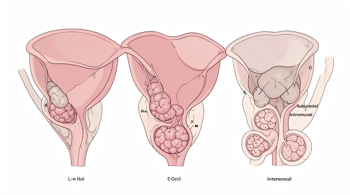
Doctors often classify fibroids based on their specific anatomical location within the uterus, which helps determine the impact on health and potential treatment pathways 25. The classification categories generally include:
- Intramural: Fibroids that grow within the muscular wall of the uterus. 26
- Submucosal: Growths that bulge inward into the uterine cavity, often affecting menstrual flow and fertility. 27
- Subserosal: Fibroids that develop on the outer surface layer of the uterine wall, typically causing external pressure on neighboring organs. 28
Clinical Management and Treatment Options
Management of uterine fibroids is highly individualized, depending on the patient's age, symptom severity, desire for future pregnancy, and overall health status 29. For those with asymptomatic or mild cases, an observational approach is frequently preferred, as many fibroids do not cause complications requiring surgical intervention 30. When medical therapy is necessary to manage heavy bleeding or discomfort, healthcare providers may utilize hormonal treatments, including oral contraceptives, progestin-only options, or levonorgestrel-releasing intrauterine devices (IUDs) 31. Non-hormonal alternatives, such as nonsteroidal anti-inflammatory drugs (NSAIDs) or tranexamic acid, may also be prescribed to address pain and acute bleeding episodes 32.
When symptoms are severe or when fertility goals are a concern, several interventional and surgical options are available. Surgical procedures like myomectomy, which involves the removal of individual fibroids while preserving the uterus, are often sought by those wishing to maintain fertility 33. Interventional radiology offers alternatives such as uterine artery embolization (UAE), which blocks the blood supply to the fibroids, causing them to shrink over time 34. Other procedures, including radiofrequency ablation and high-intensity focused ultrasound, represent additional technological advancements for symptom reduction without major open surgery 35. In instances where symptoms are debilitating and childbearing is not a priority, a hysterectomy remains a definitive surgical solution for the permanent removal of the uterus 36.
Navigating Life With Fibroids
Living with symptomatic fibroids can present daily challenges, including fatigue from blood loss and the frustration of visible abdominal distension 37. Managing these symptoms often involves a combination of medical oversight and practical lifestyle adaptations. For individuals experiencing heavy menstrual bleeding, utilizing higher-capacity menstrual products such as period underwear or menstrual cups may assist in managing flow and reducing the risk of leakage 38. Chronic pain management may require consistent collaboration with a gynecological specialist to ensure that treatments are adjusted as symptoms evolve over time 39.
Mental health support is also a recognized component of living with a chronic gynecological condition. The physical impact of fibroids can be distressing, and patients are encouraged to communicate their experiences openly with healthcare providers to address both the physical symptoms and the impact on quality of life 40. By understanding that there are diverse clinical pathways available, patients can better navigate the diagnostic and treatment processes while working toward the goal of long-term pelvic health 41.
Sources
- Yale Medicine, Uterine Fibroids (Uterine Myoma) | Fact Sheets
- National Institute of Child Health and Human Development, Uterine Fibroids
- Penn Medicine, Uterine Fibroids – Symptoms and Causes
- Loyola Medicine, Uterine fibroids: Symptoms, treatments and when to seek care
- NewYork-Presbyterian, Fibroids: 7 Facts To Know
- AskMD Sharecare, What to know about uterine fibroids
- Cleveland Clinic, Living With Uterine Fibroids
- Acibadem Hospitals Group, Fibroids: Symptoms, Causes, And Treatment
- American Family Physician, Uterine Fibroids: Rapid Evidence Review
- Nebraska Medicine, How to treat uterine fibroids
- UCLA Health, Uterine Fibroids
- USA Today, What are fibroids? Symptoms and treatment, explained
- Office on Women's Health, Uterine fibroids
- American College of Obstetricians and Gynecologists, Frequently Asked Questions: Uterine Fibroids
- Mayo Clinic, Uterine fibroids
- Johns Hopkins Medicine, Uterine Fibroids
- MedlinePlus, Uterine Fibroids
- FDA, Postmarket Drug Safety Information
- NCBI Bookshelf, Uterine Leiomyomata
- RadiologyInfo.org, Uterine Artery Embolization
- NHS, Uterine Fibroids
Authored by MyTrendSpot team