Age-related macular degeneration causes and prevention strategies: An evidence-based clinical guide
Key facts
- Age, smoking, diet, diabetes and hypertension are among factors associated with age-related macular degeneration.
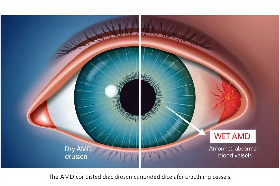
- Dry AMD involves retinal thinning and drusen, while wet AMD involves abnormal blood vessels that may leak fluid.
- Anti-VEGF treatment is delivered by injections into the eye and is mainly used for neovascular or wet AMD.
- A 2026 US cost-utility model estimated five-year direct medical costs of US$52,797 for faricimab and US$62,367 for aflibercept.
Age-related macular degeneration, commonly called AMD, affects the macula, the central retinal area responsible for detailed vision. The condition is mainly associated with ageing and can progress in different ways. Causes and prevention strategies therefore involve inherited susceptibility, smoking, cardiovascular and metabolic health, diet, eye examinations and timely specialist care. A qualified ophthalmologist or optometrist should assess suspected retinal changes rather than relying on symptoms or supplements alone.
What age-related macular degeneration is
AMD is a disorder of the macula and a major cause of irreversible central vision loss in older adults. Prevalence rises substantially with age, and a 2026 clinical review reported that approximately 19.8 million people in the United States were living with AMD in 2019, including about 1.49 million with late-stage disease. The macula supports reading, recognising faces and other detailed tasks, so damage can affect central rather than peripheral vision. 1
Most cases are classified as dry, or nonexudative, AMD. This form is associated with retinal pigment epithelial dysfunction, macular thinning and drusen, which are yellow deposits beneath the retina. Wet, or neovascular, AMD occurs when abnormal blood vessels grow beneath or near the retina and leak blood or fluid. Merck Manual reports that approximately 85% of people with AMD have only the dry form, although disease stage and progression vary between individuals. 2
Biological and environmental causes
AMD does not have one single cause. Age-related changes can impair retinal support cells, while oxidative stress, inflammation, deposits and degeneration of nearby blood vessels may contribute to damage. Genetic variants, including changes involving complement factor H, and a family history can increase susceptibility. A 2026 umbrella analysis identified age and smoking as consistent associations, while also reporting links involving diet, diabetes and hypertension, with much of the overall evidence graded low or very low quality. 3
Other reported associations include cardiovascular disease, obesity, sun exposure and diets low in omega-3 fatty acids or dark green leafy vegetables. These factors should be interpreted as contributors to risk rather than proof that one exposure directly causes AMD in a particular person. Genetic predisposition cannot be removed through lifestyle changes, and healthy habits cannot guarantee prevention. The British Journal of Ophthalmology has also examined potentially modifiable exposures, reinforcing that risk patterns are multifactorial rather than explained by one food or behaviour. 4
Risk-reduction and prevention strategies
Smoking cessation is one of the clearest modifiable measures associated with lower AMD risk, because smoking is consistently linked with the condition. Management of blood pressure, diabetes and cardiovascular health may also support general retinal and vascular health. A diet containing dark green leafy vegetables, fish and a broad range of nutrient-dense foods is commonly advised, but dietary changes should complement, not replace, eye examinations or prescribed treatment. The evidence does not support describing these measures as a cure.
Regular comprehensive eye examinations can identify drusen, pigment changes or other retinal abnormalities before noticeable central vision changes occur. An ophthalmologist may recommend monitoring at intervals based on retinal findings, visual function and the presence of AMD in one or both eyes. An Amsler grid can help with home monitoring when a clinician has recommended it, but it is not a substitute for examination. New distortion, a sudden blurred area or rapid central vision change warrants prompt professional assessment because wet AMD can progress quickly.
How AMD is diagnosed and monitored
Diagnosis generally begins with a history, visual acuity testing and a dilated examination of the retina. Ophthalmologists may use colour retinal photographs to document drusen, pigment changes or atrophy. Optical coherence tomography, or OCT, produces cross-sectional images that can show retinal swelling, fluid, thinning and structural changes. Fluorescein angiography may be used when abnormal blood-vessel leakage needs further assessment. These tests help distinguish dry changes from active neovascular disease and guide treatment decisions. 2
Monitoring is tailored to the clinical stage. Early or intermediate dry AMD may be followed through scheduled examinations, visual acuity checks, retinal imaging and symptom review. Suspected wet AMD typically requires a retina specialist because treatment decisions depend on OCT findings, leakage, visual function and previous injections. A UK hospital audit described a two-week referral-to-first-injection standard for newly referred neovascular AMD, illustrating why referral pathways can be time-sensitive, although local waiting times and service capacity differ. 5
Medical treatments for wet AMD
Anti-vascular endothelial growth factor, or anti-VEGF, medicines are injected into the vitreous, the gel-filled space inside the eye. They reduce signalling that promotes abnormal blood-vessel growth and leakage. Common treatment plans begin with repeated injections and may later use fixed or treat-and-extend intervals according to OCT findings and disease activity. Treatment is intended to control neovascular activity, not reverse every existing retinal injury. Options discussed in clinical practice include aflibercept, faricimab and ranibizumab, with selection determined by the ophthalmologist.
Intravitreal injections carry risks including eye discomfort, inflammation, bleeding on the eye surface, raised eye pressure and, rarely, serious infection inside the eye or retinal complications. Photodynamic therapy and laser photocoagulation have more limited, condition-specific roles and may be considered in selected vascular patterns. Low-vision assessment, magnification devices, lighting changes and rehabilitation can support daily function when central vision is impaired. Merck Manual lists injections, photodynamic therapy, laser treatment, dietary supplements and low-vision devices among management approaches. 2
Dry AMD supplements and clinical eligibility
Some people with intermediate AMD or advanced disease in one eye may be assessed for a specific antioxidant and mineral supplement formulation based on clinical evidence and professional guidance. Supplements are not appropriate for every stage, and some ingredients can interact with medicines or be unsuitable because of smoking history or other health factors. A retina specialist or ophthalmologist should determine whether a formulation is appropriate. Ordinary multivitamins should not be assumed to provide the same studied combination.
Eligibility for treatment depends on the AMD type, retinal imaging, visual acuity, disease activity, other eye conditions and the ability to attend repeated monitoring appointments. The risk of treatment must be weighed against the risk of untreated active disease. No supplement or lifestyle measure eliminates inherited risk, and no current strategy guarantees that dry AMD will not progress to advanced disease. Professional review is important before starting, stopping or changing supplements.
Where specialist care is provided and what it may cost
Diagnosis and treatment are usually provided by ophthalmologists, with medical retina or vitreoretinal specialists managing complicated or wet AMD. Examples of recognised specialist settings include Wills Eye Hospital in Philadelphia, where the cited professional review lists a vitreoretinal diseases and surgery service, Oslo University Hospital in Oslo, which has a Department of Ophthalmology, and Puerta de Hierro-Majadahonda University Hospital in Madrid, whose ophthalmology department has published work on neovascular AMD care. These institutions are examples for orientation, not endorsements or guarantees of access. 2 6 7
Costs depend on country, public or private coverage, medicine selected, injection frequency, imaging, consultation fees and treatment setting. A 2026 US cost-utility model estimated five-year direct medical costs of US$52,797 for faricimab and US$62,367 for aflibercept, including drug acquisition, injections, monitoring, visits and management of endophthalmitis. These are modeled healthcare costs, not a universal patient bill. Public systems may cover medically indicated care under eligibility rules, while private arrangements can involve authorisation requirements, co-payments or exclusions. 8
Sources
- Epidemiology and Risk Factors|Age-related Macular Degeneration|Clinical Guidance: https://www.healio.com/clinical-guidance/age-related-macular-degeneration/epidemiology-and-risk-factors-overview
- Age-Related Macular Degeneration (AMD or ARMD) - Ophthalmology - Merck Manual Professional Edition: https://www.merckmanuals.com/professional/eye-disorders/retinal-disorders/age-related-macular-degeneration-amd-or-armd
- Frontiers | Risk factors for age-related macular degeneration: an umbrella analysis of systematic review and meta-analysis: https://www.frontiersin.org/journals/medicine/articles/10.3389/fmed.2026.1878292/full
- Exposure-wide approaches identifying modifiable factors for age-related macular degeneration | British Journal of Ophthalmology: http://bjo.bmj.com/content/110/8/907
- NICE-informed quality improvement for neovascular age- ...: https://www.dovepress.com/nice-informed-quality-improvement-for-neovascular-age-related-macular--peer-reviewed-fulltext-article-OPTH
- Pharmacoeconomic analysis of switch to faricimab for neovascular age-related macular degeneration in real clinical practice | Global and Regional Health Technology Assessment: https://journals.aboutscience.eu/index.php/grhta/article/view/3800
- Impact of faricimab and high‐dose aflibercept on treatment demand in a tertiary ophthalmic centre in Norway - Blix - Acta Ophthalmologica - Wiley Online Library: https://onlinelibrary.wiley.com/doi/full/10.1111/aos.70240
- Cost-Utility Analysis of Faricimab Versus Aflibercept in Treating Neovascular Age-Related Macular Degeneration (nAMD) in the United States | PharmacoEconomics - Open | Springer Nature Link: https://link.springer.com/article/10.1007/s41669-026-00643-0
Authored by MyTrendSpot team