Warning signs of prolapsed uterus: Clinical indicators and diagnostic assessment pathways
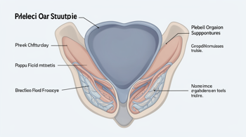
A prolapsed uterus occurs when the muscles, ligaments, and connective tissues of the pelvic floor weaken, causing the uterus to descend from its standard anatomical position into the vaginal canal 1, 2. This physiological change is a common form of pelvic organ prolapse and may occur alongside the descent of the bladder, rectum, or vaginal vault 4, 6. Understanding the early indicators of this condition is essential for patient awareness, as symptoms often manifest gradually and correlate with the severity of the descent 7, 8.
Common Clinical Manifestations and Subjective Sensations
Patients frequently report a sensation of pelvic heaviness, fullness, or a persistent dragging pressure in the lower abdominal or pelvic region 1, 9. A distinct warning sign is the feeling of tissue bulging into or protruding from the vagina, often described as a sensation that something is moving downward or coming out of the body 2, 5. Some individuals report a feeling similar to sitting on a small ball or notice vaginal tissue rubbing against clothing, particularly during periods of prolonged activity 2, 10.
These symptoms are often positional and transient, meaning they may become more pronounced after extended standing, lifting, coughing, or straining 3, 10. Conversely, many patients notice an improvement in symptoms when lying down, which reduces gravitational pressure on the pelvic floor 3. In mild cases, the physical descent may be minimal, resulting in little to no noticeable discomfort, which explains why many instances are only identified during routine pelvic examinations 2, 4.
Urinary and Bowel System Disruptions
The proximity of the uterus to the bladder and rectum means that its descent often impacts the function of these adjacent organs 1. Urinary symptoms frequently include a sense of urgency, increased frequency, or difficulty initiating urination 1, 3. Many patients describe a feeling that the bladder does not empty completely, which may lead to recurrent bladder irritation or leakage, also known as urinary incontinence 2, 3.
Bowel movements can also become complicated by the anatomical shift of the uterus. Patients may experience chronic constipation or a sense of obstruction when attempting to pass stool 3, 5. In some instances, it may become physically necessary to apply pressure against the vaginal wall with fingers to facilitate a bowel movement 2. Such mechanical changes in bodily functions are significant clinical markers that warrant professional assessment by a gynecologist or urogynecologist to determine the underlying cause and severity of the prolapse 4.
Sexual and Lumbar Comfort Considerations
Sexual dysfunction, including pain or discomfort during intercourse, is a documented concern among individuals with uterine prolapse 3, 7. Patients may report a sensation of looseness in the vaginal tissue or a physical feeling of fullness that interferes with comfortable sexual activity 2. These concerns are often tied to the anatomical changes in the vaginal canal as the cervix or uterus descends 3. Open communication with a healthcare provider can help address these functional challenges through various management strategies 4.
Furthermore, lower back pain that is not relieved by standard stretching is a recurring complaint 2, 10. While back pain can stem from numerous musculoskeletal issues, when it accompanies other pelvic symptoms, it should be evaluated as a potential indicator of pelvic floor weakness 3, 10. The persistent nature of these symptoms, even if they do not cause intense pain, should be considered as a signal to seek medical guidance regarding the status of one’s pelvic support system 9.
Risk Factors and Demographic Correlations
The prevalence of uterine prolapse is influenced by several cumulative factors that compromise the pelvic floor 1. Vaginal childbirth is a primary risk factor, particularly in cases involving a prolonged second stage of labor, the use of assisted delivery tools like vacuum extractors or forceps, or the birth of a high-birthweight infant 1, 7. Research indicates that up to 50% of women aged 50 to 79 may experience some form of this condition, with age being a notable factor in the natural loss of muscle and ligament elasticity 8.
Other critical risk factors include chronic conditions that place repeated strain on the abdominal and pelvic region, such as chronic coughing due to lung disease or frequent heavy lifting 7, 8. Obesity is also recognized as a contributor due to the increased pressure exerted on the pelvic floor over time 1, 7. Understanding these risk factors is vital for preventative efforts, which often involve lifestyle management such as maintaining a healthy weight, smoking cessation, and the management of chronic respiratory conditions 7.
Stages of Uterine Descent
Clinical staging provides a framework for healthcare providers to classify the severity of the descent and plan appropriate management 6, 7. The condition is generally categorized into degrees of advancement:
| Stage | Clinical Description |
|---|---|
| Stage 1 | The uterus has descended but remains contained within the upper vagina. |
| Stage 2 | The uterus has descended to or near the vaginal opening. |
| Stage 3 | The cervix or uterus protrudes beyond the vaginal opening. |
| Stage 4 | The entire uterus is outside the vaginal canal, representing complete prolapse. |
Diagnosis is primarily clinical, relying on a physical examination where the practitioner assesses the anatomy while the patient is asked to bear down to reveal the extent of the prolapse 1, 7. In cases of severe stage 3 or 4 prolapse, the protruding tissue may become dried, edematous, or ulcerated due to friction, which necessitates careful clinical monitoring and care to prevent infection 3.
Management and Medical Evaluation
The goal of professional evaluation is to establish an accurate diagnosis and differentiate uterine prolapse from other pelvic floor disorders 4. Conservative management strategies are typically explored first for mild cases, including targeted pelvic floor muscle exercises to strengthen support, lifestyle modifications, or the use of a vaginal pessary to provide mechanical support 1, 7. Surgical intervention is reserved for instances where symptoms are bothersome, conservative efforts prove insufficient, or the stage of the prolapse is advanced 7.
Surgical options vary depending on the patient's individual health history and may include uterine suspension, hysterectomy with vault fixation, or colpocleisis for those who do not require future vaginal intercourse 7. Since individual variation in symptoms is significant, professional consultation is required to tailor any management plan to the specific patient's needs and quality-of-life goals 7. Timely medical assessment is recommended for any individual experiencing new or worsening pelvic symptoms, bleeding, or the inability to perform normal bodily functions 6, 9.
Sources
- Merck Manual: Pelvic Organ Prolapse
- Memorial Health System: Uterine Prolapse
- Merck Manual Professional: Uterine and Apical Prolapse
- Acibadem International: Uterine Prolapse Overview
- Wikipedia: Uterine Prolapse
- Liv Hospital: Early Signs of Prolapse Uterus
- Advanced Urogynecology: Early Signs of Pelvic Organ Prolapse
- Mayo Clinic: Uterine Prolapse Symptoms and Causes
- Cleveland Clinic: Uterine Prolapse
- American College of Obstetricians and Gynecologists: Pelvic Organ Prolapse
Authored by MyTrendSpot team