Warning signs of skin cancer: A clinical guide to changes worth evaluating
Why changes in the skin matter
Skin cancer begins with abnormal growth of cells in the skin and can occur on areas that receive substantial sunlight as well as places that are usually covered. Basal cell carcinoma and squamous cell carcinoma are the most common forms and are often treatable, while melanoma is more likely to spread and can be harder to cure. A new or changing mark is therefore important to document and discuss with a healthcare professional rather than judge by appearance alone. 1
The broadest warning sign is a noticeable change in the skin. This may be a new growth, a sore that does not heal, a rough patch, or an alteration in an existing mole or freckle. Skin cancer can appear on the face, scalp, hands, feet, nails, mouth, genitals, and other areas, so a check limited to sun-exposed skin can miss relevant changes. Not every unusual spot is cancerous, but persistent or concerning changes warrant professional evaluation. 2 3
Melanoma and the ABCDE rule
The ABCDE rule is a structured way to recognize features associated with melanoma. Asymmetry means one half of a spot does not resemble the other. Border refers to an edge that is jagged or irregular. Color describes uneven shades within one lesion, including combinations of brown, black, gray, blue, red, or white. Diameter refers to a spot larger than a pea, although smaller melanomas can also require assessment. Evolving means a change in size, shape, color, or appearance over weeks or months. 2
A new spot that looks unlike a person’s other moles may also be significant, a pattern sometimes described as the “ugly duckling” sign. Melanomas can begin on normal skin or within an existing mole and may be flat, raised, irregular, or composed of several colors. They can also occur in less obvious locations, including nail beds, the mouth, eyes, and genital or rectal areas. The absence of dark pigment does not exclude concern because some melanomas appear pink, red, or skin-colored. 5 8
Signs associated with basal cell carcinoma
Basal cell carcinoma commonly develops on sun-exposed areas such as the head, neck, face, and arms, although it can occur elsewhere. A typical warning sign is a pearly, shiny, or waxy bump that may be flesh-colored, pink, or darker. Small visible blood vessels, a central depression, or a raised rolled edge can be clues. Some lesions appear as flat, pink or red scaly patches rather than bumps, so texture and persistence are relevant as well as color. 1 2
Another characteristic presentation is a sore that bleeds, crusts, oozes, or repeatedly returns after seeming to heal. Basal cell lesions may be fragile and bleed after minor irritation, shaving, or scratching. Scar-like areas that are firm, pale, yellowish, or poorly defined also deserve assessment, particularly when they arise without a clear injury. These descriptions are visual clues, not a diagnosis, because irritation, infection, and benign growths can resemble them. 3 7
Signs associated with squamous cell carcinoma
Squamous cell carcinoma often appears as a firm red or skin-colored bump, a raised growth, or a flat lesion with a rough, scaly, or crusted surface. The lesion may be painful, itchy, tender, or prone to bleeding. Open sores may ooze or form a crust, and some growths have a sunken center. These findings are especially important when they persist or enlarge instead of resolving like a short-lived rash or minor injury. 2 3
Long-term ultraviolet exposure is associated with several skin cancers, and rough scaly patches may represent actinic keratoses, which are considered precancerous lesions. A patch that feels rough when a finger passes over it, repeatedly crusts, or changes in thickness should be recorded and assessed. Squamous cell carcinoma can develop on the ears, nose, lips, hands, and other locations, but it is not restricted to visibly sun-damaged skin. 3 8
Non-healing sores and other symptoms
A sore that does not heal is one of the clearest general warning signs cited by public health guidance. Concern increases when the area bleeds, oozes, crusts, becomes tender, or heals and then returns. Mayo Clinic also identifies a new growth that resembles a mole, bump, or scab as a possible sign. The duration alone cannot confirm cancer, but an unexplained persistent lesion should not be dismissed simply because it is small. 1 2
Symptoms such as itching, tenderness, pain, tingling, or numbness can occur in suspicious lesions, although many skin cancers cause no discomfort. Some unusual presentations include scar-like pale areas, pink or red growths, and changes beneath or around a nail. A lesion that repeatedly catches on clothing, bleeds with minimal contact, or develops a different texture from surrounding skin should be included in a clinical discussion. 3 7
How to monitor the skin systematically
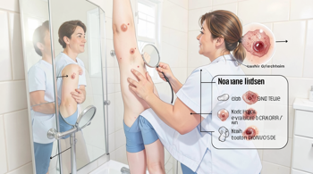
A thorough self-examination is best performed in a well-lit setting after bathing or showering, using a full-length mirror and a handheld mirror. The examination should cover the front, back, and sides of the body, including the scalp, ears, eyelids, lips, neck, underarms, hands, fingers, nails, legs, feet, soles, and spaces between the toes. A partner can help inspect the back of the scalp and other difficult-to-see areas. 4 6
Monthly checks can help establish familiarity with existing moles and make new changes easier to notice, while people with a personal or family history of skin cancer may be advised to receive examinations more often. Photographs can support comparisons of size, shape, color, and texture over time. Medical assessment is appropriate for a new growth, an evolving spot, an irregular mole, or a sore that persists, bleeds, crusts, or returns. A clinician may determine whether observation, further examination, or biopsy is appropriate. 4 6 5
Sources
- Mayo Clinic, “Skin cancer: Symptoms and causes,” https://www.mayoclinic.org/diseases-conditions/skin-cancer/symptoms-causes/syc-20377605
- Centers for Disease Control and Prevention, “Symptoms of Skin Cancer,” https://www.cdc.gov/skin-cancer/symptoms/index.html
- Penn Medicine, “Skin Cancer: Types, Symptoms, and Causes,” https://www.pennmedicine.org/conditions/skin-cancer
- UCLA Health, “Mind your moles: How to check for skin cancer,” https://www.uclahealth.org/news/article/mind-your-moles-how-check-skin-cancer
- MSD Manual Consumer Version, “Melanoma,” https://www.msdmanuals.com/home/cancer/skin-cancers/melanoma
- Banner Health, “How to Spot Skin Cancer: Warning Signs,” https://www.bannerhealth.com/healthcareblog/teach-me/how-to-spot-skin-cancer
- Cleveland Clinic, “Nonmelanoma Skin Cancer: Symptoms, Types & Treatment,” https://my.clevelandclinic.org/health/diseases/nonmelanoma-skin-cancer
- American Academy of Dermatology, “ABCDEs of Melanoma,” https://www.aad.org/public/diseases/skin-cancer/find/at-risk/abcdes
Authored by MyTrendSpot team