Warning signs of uterine fibroids: Clinical indicators and diagnostic considerations
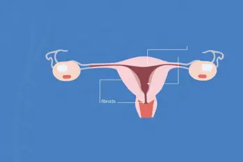
Uterine fibroids, scientifically referred to as leiomyomas or myomas, represent the most frequent type of benign tumor encountered in women of reproductive age 179. While these growths of smooth muscle and fibrous tissue are pervasive, affecting up to 70% to 80% of women by the age of 50, their clinical manifestation is highly variable 147. Many individuals remain entirely asymptomatic, with these growths often discovered incidentally during routine pelvic examinations or obstetric ultrasounds 578. When symptoms do occur, they are typically dictated by the size, number, and precise anatomical location of the fibroids within or around the uterus 256.
Understanding the Symptom Profile
The most commonly reported clinical sign of uterine fibroids is abnormal uterine bleeding, specifically characterized by heavy or prolonged menstrual cycles lasting seven days or more 256. This persistent blood loss can lead to secondary health complications, such as anemia 16. Beyond menstruation, individuals frequently report pelvic pressure or pain, which may manifest as a persistent sensation of heaviness in the lower abdominal region 256. Because these symptoms can overlap with other gynecological conditions, clinical evaluation is essential for accurate identification 49.
Bladder and Bowel Pressure Indicators
As fibroids enlarge, they can exert mechanical pressure on adjacent pelvic organs, leading to distinct physiological changes 12. If a fibroid presses against the bladder, common indicators include increased urinary frequency or a persistent sense of urgency, as well as difficulty in fully emptying the bladder 256. Similarly, if the growths compress the rectum, patients may experience chronic constipation or noticeable rectal pressure 245. These bulk symptoms are often among the primary drivers for patients to seek formal medical consultation 49.
Structural and Reproductive Manifestations
Large or rapidly growing fibroids can lead to visible abdominal distension, where the enlargement of the uterus becomes significant enough to alter the patient's physical silhouette, occasionally mimicking the appearance of pregnancy 257. Furthermore, internal pressure can radiate to other parts of the body; lower back pain and localized leg pains are sometimes reported when fibroids impinge upon the pelvic nerves 25. In the context of sexual health, some women report experiencing discomfort or pain during intercourse, known clinically as dyspareunia, which is heavily dependent on the positioning of the fibroids 256.
Diagnostic Approaches and Clinical Evaluation
Medical professionals generally begin the diagnostic process with a comprehensive gynecological history, followed by abdominal, speculum, and bimanual pelvic examinations 9. For definitive identification, the standard initial imaging modality involves a combination of transvaginal and transabdominal ultrasonography 9. In more complex cases, or when the extent of the disease is unclear, pelvic magnetic resonance imaging (MRI) with contrast may be employed to better characterize the location and size of the growths 9. These diagnostic tools are critical in distinguishing fibroids from other reproductive system pathologies 4.
Risk Factors and Demographic Trends
While the exact etiology of uterine fibroids remains partially unclear, specific demographic and biological risk factors have been established through clinical research. Being of reproductive age is the primary risk factor, with African American women demonstrating a higher prevalence and a tendency to develop these growths at younger ages 13. Genetics also play a significant role, as a positive family history increases an individual's likelihood of development 134. Furthermore, the growth of these tumors is hormonally mediated; specifically, estrogen and progesterone levels are known to stimulate development, explaining why fibroids often shrink following menopause 234.
Management Philosophy and When to Seek Care
Medical intervention is not required for every case of uterine fibroids, as many remain small and asymptomatic throughout a patient's life 13. However, when symptoms interfere with daily comfort, blood levels, or reproductive goals, various clinical pathways are available 69. Treatment strategies range from watchful monitoring and hormonal pharmacotherapy to minimally invasive procedures and surgical options 169. Patients experiencing severe, sudden pain, fainting, or signs of extreme blood loss are advised to seek immediate medical review to rule out secondary complications 6.
Sources
- Yale Medicine: Uterine Fibroids (Uterine Myoma) Fact Sheet
- Penn Medicine: Uterine Fibroids Symptoms and Causes
- Loyola Medicine: Uterine Fibroids Symptoms, Treatment, and Care
- NewYork-Presbyterian: How Can I Tell if I Have Fibroids?
- Memorial Health System: Uterine Fibroids Health Library
- Acibadem Hospitals Group: When To See A Doctor For Uterine Fibroids
- Manipal Hospitals: Uterine Fibroids Explained: A Woman’s Guide
- Cleveland Clinic: 5 Tips for Living With Uterine Fibroids
- American Family Physician: Uterine Fibroids Rapid Evidence Review
- Lona Sasser Obstetrics & Gynecology: Uterine Fibroid Symptoms
Authored by MyTrendSpot team