Glaucoma Early Detection: A Comprehensive Review of Diagnostic Innovations and Artificial Intelligence Applications
Glaucoma early detection remains a critical objective in global ophthalmic health because the disease typically presents no symptoms in its initial stages. Clinical data indicates that more than 3 million people in the United States currently have glaucoma, yet approximately 50% are entirely unaware of their condition 1. This asymptomatic progression often leads to irreversible optic nerve damage before functional vision loss is noticed by the patient. Research from the National Eye Institute confirms that identifying structural changes early is the only viable pathway to preventing permanent blindness 2. Since the disease is characterized as a progressive optic neuropathy, the focus of modern medicine has shifted toward high-precision structural mapping and risk-based screening protocols 3.
Artificial Intelligence Systems in Clinical Environments
The integration of artificial intelligence (AI) into clinical environments has significantly altered the landscape of glaucoma screening. A retrospective cross-sectional study evaluated the diagnostic accuracy of two systems, Laguna ONhE and VUNO Med-Fundus AI, using color fundus photography (CFP). The results showed that Laguna achieved an area under the receiver operating characteristic curve (AUC) of 0.879, while VUNO showed an AUC of 0.857. When these two systems were utilized in combination, the diagnostic accuracy improved to an AUC of 0.903, which is statistically comparable to traditional global mean deviation metrics 4. This suggests that AI can serve as a robust alternative in settings where more expensive equipment is unavailable.
Further advancements include the development of Vision Transformer (ViT) models, which analyze optic disc photographs to classify eyes as glaucomatous or healthy. In one test phase involving over 1,400 eyes, a ViT model achieved an overall accuracy of 0.987 with a sensitivity of 0.994 and a specificity of 0.969 5. Another model, GlaucoXAI, utilizes grey-wolf optimization and extreme learning machines to reduce model overfitting. This system demonstrated accuracy rates of 93.87% and 95.38% across benchmark datasets, providing clinicians with interpretable diagnostic data that enhances trust in automated results 6.
Structural Analysis through Optical Coherence Tomography
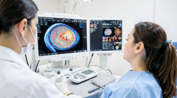
Optical Coherence Tomography (OCT) has become a standard tool for detecting structural damage that precedes functional vision loss. A multidimensional imaging model was recently developed to combine fundus photographs with retinal nerve fiber layer (RNFL) and ganglion cell-inner plexiform layer (GCIPL) thickness maps. This integrated approach achieved a superior AUC of 0.970, outperforming models that relied solely on fundus photography or individual structural maps 7. Such data highlights the necessity of multimodal structural analysis in identifying primary open-angle glaucoma (POAG) at the earliest stages.
| Metric Analyzed | Traditional Fundus Model (AUC) | Multidimensional Model (AUC) |
|---|---|---|
| RNFL and GCIPL Deviation | 0.945 | 0.970 |
| Ganglion Cell Thickness | 0.915 | 0.970 |
| Optic Nerve Head Metrics | 0.890 | 0.970 |
A prospective longitudinal cohort study followed 244 eyes for an average of 30.8 months to determine the lead time advantage of OCT in detecting disease progression. The research found that serial spectral-domain OCT measurements of peripapillary RNFL and macular ganglion cell layers can identify progressive thinning before measurable functional loss occurs in visual field testing 8. Furthermore, routine screening using OCT has been shown to identify glaucoma suspects who would not have been referred for further evaluation during standard eye examinations, although researchers emphasize that the rate of false positives and the associated costs must be carefully considered 9.
Field Testing and Mobile Screening Technologies
In regions where access to hospital-grade equipment is limited, portable and mobile screening technologies are filling a critical diagnostic gap. A study conducted in Nigeria compared portable devices against standard reference tools, finding strong correlations between iCare tonometry and Goldmann Applanation Tonometry (GAT) (r = 0.96) 10. The Remedio handheld fundus camera achieved a high diagnostic accuracy with an AUC of 0.91 and a specificity of 99.8%. These portable tools are often faster to use, have higher completion rates, and are strongly preferred by participants over conventional clinical machinery 10.
Smartphone-based diagnostic tools are also emerging as highly efficient options for community screening. Doctors at the LV Prasad Eye Institute developed an AI-powered smartphone tool that detects glaucoma with 92.02% accuracy. For early-stage cases specifically, the tool maintained an accuracy of 86.9% 11. Complementing these structural tools are tablet-based visual field checkers, such as the iPad Quattro Checker (iPad QC). This device retained 80.2% sensitivity in early glaucoma and helped over half of previously unaware patients recognize their visual field deficits during a self-administered test lasting only a few minutes 12.
Demographic Disparities and Risk-Based Evaluation
Research indicates that certain demographic groups are disproportionately affected by glaucoma, necessitating equity-aware screening models. African ancestry individuals are often under-represented in medical datasets, yet a deep learning model developed specifically for this population achieved an AUC of 0.925 in screening for POAG 13. In parallel, the FairDist model was proposed to utilize baseline OCT scans while ensuring equal diagnostic outcomes across different gender and racial groups, achieving high equity-scaled AUC scores 14. This focus on fairness is intended to mitigate disparities in disease progression prediction.
- Individual Risk Factors: Adults over the age of 60 and those with a family history are at significantly higher risk for development 15.
- Ethnic Variations: Individuals of African, Hispanic, or Asian descent show higher prevalence rates of specific glaucoma types
- Myopic Considerations: Deep learning autoencoders have been specialized for detecting glaucoma in myopic eyes, which often present unique structural challenges 16.
- Geographic Prevalence: In Asian populations, the prevalence of primary angle-closure glaucoma ranges from 0.75% to 1.19%, requiring specialized screening for narrow anterior chamber angles 17.
Regulatory Perspectives and Implementation Barriers
Despite the proliferation of diagnostic technology, significant barriers to universal screening persist. In Australia, between 50% and 60% of cases remain undiagnosed, contributing to an economic impact projected to reach $4.3 billion by 2025 in healthcare costs and lost wellbeing 18. The U.S. Preventive Services Task Force (USPSTF) currently maintains that there is insufficient evidence to fully assess the balance of benefits and harms of universal screening for open-angle glaucoma in adults 19. This creates a landscape where opportunistic screening during routine visits is the primary method for detection.
Primary care settings often face challenges such as inconsistent screening protocols and limited access to specialized equipment like slit lamps or Humphrey field analyzers. Scoping reviews indicate that most glaucoma screening is currently performed at the secondary healthcare level by eye specialists, rather than at the primary care level 20. To address this, organizations like the American Academy of Ophthalmology recommend that all adults receive a baseline eye screening by age 40 21. Effective management and prevention of vision loss ultimately depend on the coordination of care between general practitioners, optometrists, and ophthalmologists to ensure timely referrals 18.
Sources
- Centers for Disease Control and Prevention (CDC)
- National Eye Institute (NIH)
- American Academy of Ophthalmology (AAO)
- PLOS One Journal: Combined diagnostic accuracy of two AI systems
- Scientific Reports: Vision transformer model for glaucoma detection
- PLOS Computational Biology: GlaucoXAI diagnosis model
- BMJ Open Ophthalmology: Multidimensional imaging model study
- CME Journal Geriatric Medicine: OCT-guided progression study
- Healio Optometry: OCT during routine eye exams study
- Eye Journal: Comparison of portable devices in LMICs
- Telangana Today: LV Prasad Eye Institute AI-smartphone tool
- Graefe s Archive for Clinical and Experimental Ophthalmology: iPad Quattro Checker study
- npj Digital Medicine: Deep learning model for African ancestry individuals
- npj Digital Medicine: Equity-enhanced glaucoma progression prediction
- Glaucoma Research Foundation
- Frontiers in Ophthalmology: Glaucoma detection in myopic eyes
- npj Digital Medicine: Screening based on portable slit lamp
- Royal Australian College of General Practitioners (RACGP)
- U.S. Preventive Services Task Force (USPSTF)
- F1000Research: Challenges in primary health care level screening
- American Academy of Ophthalmology: Early detection recommendations
Authored by MyTrendSpot team